Pain
Diagnosing Crohn’s Disease

What is Crohn’s disease?
Crohn's disease is an inflammatory bowel disease (IBD) that causes inflammation of the digestive tract. Although the intestines are usually the most symptomatic, Crohn’s disease can produce symptoms anywhere in the digestive tract from the mouth to the anus. The lower part of the small intestine is most commonly affected by the disease. Symptoms include, but are not limited to, stomach pain, diarrhea, blood in the stool, fatigue, and weight loss.
How is Crohn’s disease diagnosed?
Diagnosing Crohn’s disease typically begins with a visit to a primary care physician or a gastroenterologist. The diagnostic process involves obtaining a detailed personal medical history, family health history, and a physical exam. To confirm a suspected diagnosis, blood tests, imaging tests, a stool study, a colonoscopy, or an endoscopy may be ordered.
Blood tests
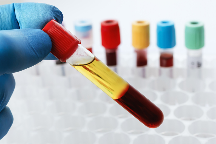
Blood tests that may be performed during the diagnostic process include the following:
- Complete blood count (CBC)
A CBC can identify low red blood cell counts (anemia) and signs of inflammation, which can indicate Crohn’s disease. Approximately one-third of people with Crohn’s disease have anemia. - Antibody tests
Blood tests that check for the anti-saccharomyces cerevisiae antibody or the perinuclear anti-neutrophil cytoplasm antibody can help in the diagnosis of Crohn’s disease or ulcerative colitis. - C-reactive protein level
This is another blood test that can identify inflammation in the body. - Iron and B12 level tests
Levels of iron and B12 may be low if Crohn’s disease prevents the small intestine from absorbing vitamins and minerals like it should.
Imaging studies
- Computerized tomography (CT) scan
A CT scan creates images of the digestive tract. It typically shows inflammation in the intestines if Crohn’s disease is present. - Magnetic resonance imaging (MRI)
An MRI uses magnetic fields and radio waves to create images of tissues and organs. It is especially helpful in allowing doctors to see an anal abscess or fistula. - Barium X-ray
Before these X-rays are taken, a chalky substance containing barium is given by mouth or through the rectum. The substance flows through the digestive tract and shows up on X-rays, allowing physicians to see ulcers, narrowed areas in the intestines, or other issues.
Other tests
- Stool study
A stool sample may be collected to check for bacteria or parasites. It can be used to eliminate the possibility of other causes of symptoms (e.g., diarrhea). - Colonoscopy
A colonoscopy is the most commonly used procedure to diagnose Crohn’s disease. During a colonoscopy, a physician uses a thin, flexible tube with a light and camera to view the inner walls of the colon. The tool can also remove small tissue samples for testing. The presence of inflammatory cells known as granulomas suggests Crohn’s disease. - Upper gastrointestinal (GI) endoscopy
During an upper GI endoscopy, a physician threads a thin, flexible tube with a light and camera down the throat to view the inner walls of the esophagus, stomach, and upper intestines. The endoscope can also remove small tissue samples for testing.
Although there is not one specific test for Crohn’s disease, these tests can eliminate the possibility of other conditions and determine a proper diagnosis so that treatment can begin.


